From FlyWire to FLY-002: Rebuilding a Fly Brain in the Browser
What FlyWire scientists mapped, why FLY-001 continued into the more complete FLY-002, and where full-graph simulation still ends.
In September 2026 I set myself a deliberately uncomfortable engineering question: if a complete fly connectome is public, how far can I take it inside an ordinary website before the words “digital brain” become misleading? FLY-001 made the question tangible, but it also exposed where a reduced graph, generic dynamics and a procedural body were too easy to overread. We continued to FLY-002 to make the experiment more complete, more measurable and more honest about what comes from data and what comes from code.
This article begins with the science because the distinction matters. FlyWire is a landmark anatomical reconstruction. My project is an experimental software interpretation built on top of a derived dataset. It is not a preserved animal, a verified copy of a fly mind, or evidence of consciousness in a browser.
First: what the FlyWire researchers actually mapped
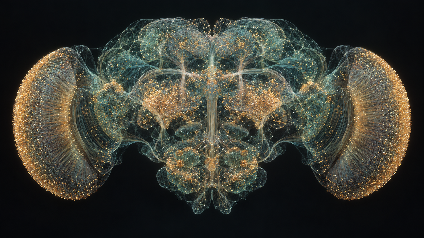
The FlyWire Consortium reconstructed the brain of one adult female Drosophila melanogaster from serial-section electron microscopy. Automated segmentation turned the enormous image volume into candidate cells; researchers, professional proofreaders and community contributors then corrected and annotated the reconstruction. The flagship Nature wiring-diagram paper reports 139,255 proofread neurons and 54.5 million synapses across the central brain and both optic lobes.
Those two numbers describe different levels of the graph. A synapse is an individual contact, while a directed neuron pair can contain many contacts. The public FAFB v783 dataset in FlyWire Codex therefore exposes neuron, annotation, synapse and connection tables rather than one universal “connection count.” FLY-002 rebuilds the released table without a strength cutoff: 16,847,997 pair-by-neuropil rows become 15,091,983 directed neuron pairs while retaining all 54,492,922 published synapse counts. Zero source rows are intentionally omitted.
The companion whole-brain annotation study made the geometry searchable and biologically legible. It organized neurons into hierarchical classes and 8,453 annotated cell types, with information about sensory, intrinsic, ascending, descending, motor and other systems. Without that work, a connectome is an extraordinary graph but a difficult one to address from code: inputs, candidate integrators and outputs are not self-explanatory from node IDs alone.
A connectome is a wiring diagram. It records anatomical relationships; it does not record the living animal’s moment-to-moment voltages, synaptic chemistry, hormones, learning history, body state or environment.
Why a wiring diagram is not yet a working brain
Connectivity strongly constrains what a circuit can do, but it does not uniquely specify its dynamics. Real neurons differ in membrane properties, morphology and electrical compartmentalization. Synapses have signs, delays, receptor combinations, nonlinearities and plasticity. Many early visual neurons in Drosophila communicate with graded potentials rather than clean all-or-nothing spikes. Glia and neuromodulators change the operating regime. Most of those parameters cannot be read directly from an electron-microscopy reconstruction.
There is encouraging evidence that structure can still be an unusually powerful constraint. A 2024 connectome-constrained visual-system model used measured connectivity for 64 optic-lobe cell types, optimized unknown cellular and synaptic parameters for motion estimation, and produced neural-response predictions consistent with measurements reported across 26 experimental studies. That is a much stronger scientific protocol than simply assigning the same equation to every cell: connectivity was combined with a defined task, fitted parameters and external validation.
Embodiment adds another layer. NeuroMechFly v2 couples a data-driven fly body, sensors, muscles, neural controllers and physics. Its lesson is simple: behavior belongs to a closed loop between nervous system, body and world. A brain graph floating in isolation cannot tell us how wing aerodynamics, proprioception, collisions or energy constraints shape action.
The question I chose to test
I was not trying to reproduce all of that biology. I wanted to test a narrower product and systems question: can the released neuron set and connectivity table be made interactive in a normal browser, attached to a visible body, given one identity across visitors, and instrumented well enough that a curious person can separate model output from software choreography?
FLY-001 began with the MIT-licensed snedea/flybrain project and proved that 139,255 neuron records could run in the portfolio. We continued to FLY-002 because the first runtime used a reduced set of 2,698,236 weighted connections. For FLY-002 I rebuilt the asset from the pinned FAFB v783.0 release, preserved every canonical uint64 neuron ID and every positive synapse count, and aggregated only repeated regional rows belonging to the same directed neuron pair. A manifest records the source checksum, generated hash, counts and limitations.
Inside the browser brain
The FLY-002 neural runtime lives in a Web Worker, away from React rendering and the 3D scene. Its compressed full graph is about 47.5 MB and decodes into a compact CSR representation of about 92.8 MB. The dynamics are based on the published whole-brain LIF model by Shiu et al.: a 0.2 ms integration step, 1.8 ms synaptic delay, 2.2 ms refractory period and transmitter-aware excitation or inhibition. Each 100 ms wall-clock update advances 25 model steps, or 5 ms of simulated time—roughly 0.05× biological time when the browser keeps up.
Sparse active-cell integration carries voltage, conductance, refractory state and delayed spikes without scanning or copying the entire graph on every small step. Acetylcholine is treated as excitatory; GABA and glutamate as inhibitory. Dopamine, serotonin, octopamine and unknown predictions are simplified as fast excitation, including 19,658 cells with unknown transmitter labels. Those choices are explicit model assumptions, not receptor-level reconstruction.
Light, temperature, touch and air are translated into seeded sensory drives whose geometry and gain remain heuristic. The first version taught an important lesson: a worker can keep ticking while a poorly chosen input produces no meaningful response. In FLY-002 the tests therefore check real propagation, non-zero sensory and reward spikes, delays, refractory behavior and activity accumulated between interface frames—not merely a moving counter.
Giving FLY-002 a body, a room and something to do
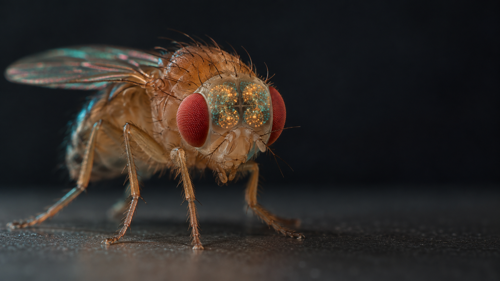
FLY-002 appears inside the interactive Baku studio using a body derived from the microscopy-based female model in FlyBody. It is a different specimen from the FlyWire brain, enlarged roughly 65× so visitors can inspect it. Its naturalistic materials and wing animation improve embodiment, but the body is not a one-to-one match and the wing motion is illustrative rather than a MuJoCo neuromechanical simulation.
Autonomous flight stays inside the room and blends exploration, phototaxis, startle, feeding and rest. Visitors can inspect the body, follow it, see through a fly-level camera, or override heading, altitude and speed while the brain keeps running. The first-person view is cinematic, not compound-eye optics. The neural map renders 10,001 sampled cell coordinates to keep the display practical; all 139,255 model neurons continue to run.
To turn observation into measurable interaction, I built a shelf-sized airfield and a seven-gate course. A visitor can fly a miniature plane, chase FLY-002 and fire a nonlethal air pulse. Valid hits stimulate the mapped Johnston’s-organ group and provoke escape steering; ordered ring passes can stimulate a reward group. The visit-local gate preference changes guided speed, but the FlyWire weights never change. This is a transparent game-learning rule inspired by dopamine-mediated reinforcement research, not evidence that the reconstructed brain learned like the source animal.
One identity, many visitors
I wanted FLY-002 to be one named organism rather than a fresh animation in every tab. Protocol version 2 and a separate Durable Object identity prevent an old FLY-001 coordinator or checkpoint from being presented as the new graph. Exactly one connected browser becomes leader and runs the neural worker; other visitors observe the canonical world state. The leader publishes validated movement and periodically uploads a checkpoint containing voltage, conductance, refractory state, delayed spikes, random-generator state and simulation time. If it disconnects, leadership can move to another client.
There is an important operational caveat. Durable Object hibernation can preserve identity and world state without billing a permanently active process, but it cannot execute every neural tick while nobody is connected. An optional headless Chromium caretaker can keep the loop alive. Until the coordinator and caretaker are deployed, the public interface can fall back to a clearly labelled per-browser simulation. Persistence is an implemented architecture with deployment requirements, not magic permanence supplied by the connectome.
A neural journal, not a mind reader
The compact FLY-002 monitor grows when a visitor wants detail. It reports model time, compute time, behavior, speed, height, drives and complete twenty-update activity windows. Those windows count every spike and every distinct firing cell instead of sampling a single convenient frame. The downloadable JSON journal and identity-preserving 10,001-point map make quiet periods, repeated firing, reward responses and air-pulse responses inspectable without inventing activity for visual effect.
Gemini 3.1 Flash-Lite can translate one validated telemetry snapshot into a short sentence. The request contains no conversation history, image or connectome data, uses minimal reasoning, is limited to 80 output tokens and is capped by minute, page-session and server budgets. The UI calls the result an AI interpretation. The model summarizes fields such as “startle,” “speed” and “fired neurons”; it does not decode experience or report what a fly literally thinks.
Real connectome data + simulated neural dynamics + heuristic embodiment + AI-written telemetry is still a simulation. Each “+” adds capability, not proof of consciousness.
What the experiment demonstrates
Technically, FLY-002 shows that the complete released connectivity table can be packaged for a website, advanced off the main thread, checkpointed, connected to an interactive 3D scene and inspected on desktop and touch devices. On this workstation the validated worker initialized in about 63 ms and averaged about 22 ms per 100 ms update, although that is not a phone guarantee. The full graph contains 5.59× as many directed pairs as FLY-001, so the improved fidelity deliberately costs more CPU even after removing runtime sorting, graph duplication and float-weight expansion.
Scientifically, it demonstrates much less—and that boundary matters. The dynamics are paper-inspired but are not validated as equivalent to recorded whole-animal responses. Synapse locations, receptor combinations, neuromodulation, glia, memories and the ventral nerve cord are missing. The body belongs to another specimen; the plane uses assisted indoor-game physics; sensory geometry, drives and reward policy are approximate. No behavior should be treated as a prediction about the animal whose brain was imaged.
Where I would take it next
Continuing beyond FLY-002 should mean calibration rather than a larger spectacle: compare responses with published recordings, introduce cell-type and receptor-specific dynamics, connect the brain to ventral-nerve-cord data and a validated neuromechanical body, and define tasks whose failure can be measured. Deploying the version-2 coordinator and caretaker would also turn the current local identity architecture into a persistent public organism without pretending that persistence is consciousness.
The fly connectome is extraordinary because it lets us ask these questions with concrete anatomy instead of a generic neural network. FLY-001 made that anatomy interactive; we continued to FLY-002 to preserve far more of the released graph, use a stronger dynamics model and make each claim inspectable. It is close enough to feel alive and transparent enough to show why “alive” remains a metaphor.
Sources and further reading
Dorkenwald et al., Neuronal wiring diagram of an adult brain, Nature (2024)
Wang-Chen et al., NeuroMechFly v2, Nature Methods (2024)
FlyWire Codex — FAFB v783 data explorer
snedea/flybrain — the MIT-licensed browser simulator adapted for this experiment
FlyWire FAFB v783.0 released connectivity table
Shiu et al., A Drosophila computational brain model, Nature (2024)